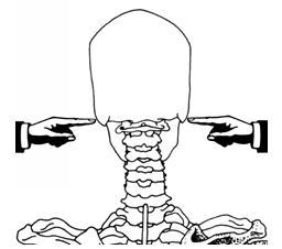
The spinal column is separated into 5 sections. Seen from the side, a human’s healthy spine has two curves resembling the shape of the letter “S”.
The cervical (neck) (red color) / C 1-7 has an inward curve. Arteries that carry blood to the brain pass through openings in the side of the cervical vertebrae. The flexible neck supports the head, which is like a bowling ball. The head balances on the first cervical vertebra called the atlas and pivots on the second vertebra called the axis.
A head neck joint has two joints. The atlanto-occipital joint and the atlanto-axial joint.
Atlas (C1), and Axis (C2) vertebrae
Knowing how the cervical vertebrae function is important to understand how your head and neck moves and stays in proper alignment.
This site provides an exercise to help you find the pivot point of your head on your spine. http://www.alexandertechniqueinoxford.com/critical-for-the-alexander-technique-finding-the-top-of-the-spine/
The thoracic (chest) (blue color) / T 1 – 12 has an outward curve. This area of the spine is very stable. The vertebrae are attached firmly to the ribs and sternum (breast bone).
Lumbar (abdominal & lower back) (yellow color) / L 1 – 5 has an inward curve. These vertebrae are able to move and flex. Sometimes people are born with a sixth vertebra in the lumbar region
Sacral (buttocks) (green color) / S1-5 fused into one has an outward curve. They are at the base of the spine and form part of the pelvic area.
Coccyx (tail bone) (purple color) / fused


Edit post: Remove video that no longer functions. Replaced with video showing skeletal C1 and C2 vertebrae and how they sit together. This isn’t as good a video as prior one for demonstrating neck joint movement.